Home » Uncategories » Left Hip Muscles Anatomy : Why Does The Outside Of My Hip Hurt What To Do About It Lakeview Physiotherapy Blog / There are 3 main layers of hip abductor muscles:
Left Hip Muscles Anatomy : Why Does The Outside Of My Hip Hurt What To Do About It Lakeview Physiotherapy Blog / There are 3 main layers of hip abductor muscles:
Left Hip Muscles Anatomy : Why Does The Outside Of My Hip Hurt What To Do About It Lakeview Physiotherapy Blog / There are 3 main layers of hip abductor muscles:. Ligaments, tendons, and muscles play an important role in the function of the hip. Let the left knee fall outward as much as possible. The posterior muscle group is made up of the muscles that extend (straighten) the thigh at the hip. You can strain or tear your hip flexor muscles through sudden movements or falls. Anatomy of the hip joint muscles | medicinebtg.com :
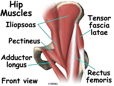
Anatomy of the hip muscles. You can strain or tear your hip flexor muscles through sudden movements or falls. Lateral rotation is needed for crossing the legs. Posterior view of gluteus maximus and gluteus medius in human anatomy, the muscles of the hip joint are those muscles that cause movement in the hip. These are gracilis, pectineus, adductor longus, adductor brevis, adductor magnus, and adductor minimus muscles.
Hip Anatomy Eorthopod Com from eorthopod.com The femur may also rotate around its axis about 90 degrees at the hip. Rectus femoris muscle, one of. These muscles are responsible for hip joint extension (backward movement). The gluteals make up the muscles of the buttocks on the back of the hip. Lateral rotation is needed for crossing the legs. The muscles of the neck can be divided into groups according to. Your body has two iliopsoas muscles: The hamstrings are three muscles at the back of the thigh that affect hip and knee.
Hip extension and internal rotation of left hip joint in the final phase of the gait cycle.
Extensors are located on the back side of the thigh. The femur may also rotate around its axis about 90 degrees at the hip. You can strain or tear your hip flexor muscles through sudden movements or falls. Attached to the bones of the skeletal system are about 700 named. Anterior muscles extend your legs and flex your thighs. The gluteals make up the muscles of the buttocks on the back of the hip. The hamstrings are three muscles at the back of the thigh that affect hip and knee. They are important for stabilising the body and for moving the legs. Hip muscle anatomy is a complex topic. Related posts of muscles of the lower back and hip diagram human muscle anatomy neck. Here we explain the hip and groin muscles, their actions and exercises. The hip muscles include pelvic and groin muscles. Muscle movements, types, and names.
Your email address will not be published. Posterior view of gluteus maximus and gluteus medius in human anatomy, the muscles of the hip joint are those muscles that cause movement in the hip. These muscles include the gluteus maximus muscle (the largest muscle in the body) and the hamstrings group, which consists of the biceps femoris, semimembranosus, and semitendinosus muscles. Human muscle anatomy neck 12 photos of the human muscle anatomy neck human anatomy muscles of the neck, human muscle anatomy neck, muscle anatomy neck and shoulder, human muscles, human anatomy muscles of the neck, human muscle anatomy neck, muscle anatomy. The sartorius muscle is a distinctively long and thin muscle that crosses the thigh diagonally.
Assessment Of The Young Adult Hip Joint Using Plain Radiographs Springerlink from media.springernature.com Muscle movements, types, and names. The muscles that flex the hip are in front of the hip joint. Functionally, the hip joint enjoys a very high range of motion. These muscles include the gluteus maximus muscle (the largest muscle in the body) and the hamstrings group, which consists of the biceps femoris, semimembranosus, and semitendinosus muscles. These muscles are the adductor longus, adductor brevis, adductor magnus, gracilis, and the obturator externus. Let the left knee fall outward as much as possible. Here we explain the hip and groin muscles, their actions and exercises. There are 3 main layers of hip abductor muscles:
Blood vessels and nerves of the hip
One at the left hip, and one at the right hip. These muscles include the gluteus maximus, hamstrings muscle group consisting of the biceps femoris, semimembranosus, and semitendinosus, and the adductor magnus. Attached to the bones of the skeletal system are about 700 named. There are also diseases and disorders that can cause the pain to. They allow you to move your leg or knee up towards your torso, as well as to bend your torso forward at the hip. The iliofemoral, pubofemoral, and ischiofemoral ligaments represent the thickenings of the joint capsule. Iliopsoas muscle, a hip flexor muscle that attaches to the upper thigh bone. Learn about hip muscles human anatomy with free interactive flashcards. Here we explain the hip and groin muscles, their actions and exercises. Want to learn more about the anatomy of the hip adductors? This mri hip joint axial cross sectional anatomy tool is absolutely free to use. These muscles are responsible for hip joint extension (backward movement). These ligaments reinforce and stabilize the hip joint(6).
If soft tissue, such as skin, muscles, fat, and fascia get strained or injured, left hip pain can come from the abdominal wall. These muscles include the gluteus maximus, hamstrings muscle group consisting of the biceps femoris, semimembranosus, and semitendinosus, and the adductor magnus. This tutorial will teach you all about the six hip adductor muscles. Use the mouse scroll wheel to move the images up and down alternatively use the tiny arrows (>>) on both side of the image to move the images.>>) on both side of the image to move the images. The hip's essential muscles are the sartorius, rectus femoris, gluteus minimus and medius, iliopsoas, adductors, and hamstrings.
Pin On Health Fitness from i.pinimg.com Posterior view of gluteus maximus and gluteus medius in human anatomy, the muscles of the hip joint are those muscles that cause movement in the hip. One at the left hip, and one at the right hip. It is also referred to as a ball and socket joint and is surrounded by muscles, ligaments, and tendons. Blood vessels and nerves of the hip Hip flexors (femoral n.) anatomy. Injury to the iliopsoas may cause hip pain and limited mobility. The quadriceps group of four muscles. Iliopsoas muscle, a hip flexor muscle that attaches to the upper thigh bone.
These muscles are responsible for hip joint extension (backward movement).
The main action of the adductors is to pull the leg inward toward the other leg. Hip flexors (femoral n.) anatomy. The hip muscles include pelvic and groin muscles. The hip muscles are composed of multiple flexors, extensors, adductors, abductors, and rotators that work together. Pelvis and acetabulum, with muscle attachment sites. Your email address will not be published. The view on the left has the rectus femoris cut away to show the vastus intermedius which is below it. Attached to the bones of the skeletal system are about 700 named. The six hip adductor muscles are all located in the adductor or medial compartment of the thigh and all mainly adduct the thigh at the hip joint. The hamstrings are three muscles at the back of the thigh that affect hip and knee. You can strain or tear your hip flexor muscles through sudden movements or falls. It is also referred to as a ball and socket joint and is surrounded by muscles, ligaments, and tendons. The iliofemoral, pubofemoral, and ischiofemoral ligaments represent the thickenings of the joint capsule.
0 Response to "Left Hip Muscles Anatomy : Why Does The Outside Of My Hip Hurt What To Do About It Lakeview Physiotherapy Blog / There are 3 main layers of hip abductor muscles:"


0 Response to "Left Hip Muscles Anatomy : Why Does The Outside Of My Hip Hurt What To Do About It Lakeview Physiotherapy Blog / There are 3 main layers of hip abductor muscles:"
Post a Comment